Originally published in Plant Healer Quarterly
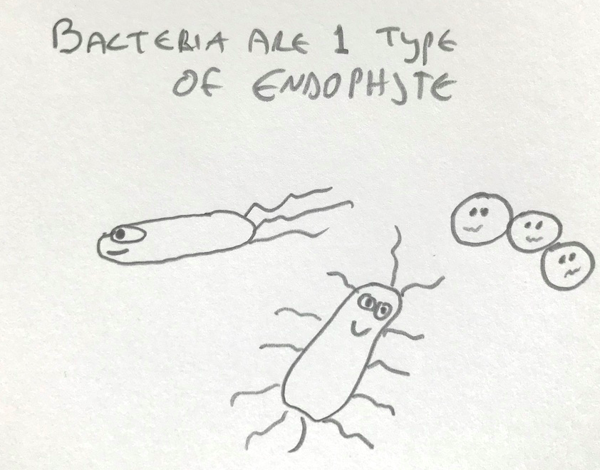
Endophytes are little critters that live either inside of or squished in between plant cells, and they’re a reminder that no organism, whether plant, person or platypus, is an individual. Rather, we’re communities of our own cells along with enormous numbers of resident microbes. We even have microbial DNA integrated into our chromosomes. And these bacterial, fungal and other critters themselves have their own collection of even smaller critters, namely viruses, within them. Fractal nature…as above, so below.
It’s clear that these little tag-alongs — endophytes, the gut microbiome, etc — have a big impact on health in us and in plants, and most likely in platypuses as well. (By the way, did you know that the correct plural form is actually platypodes? Who knew??). “Endophyte” the word came from a 19th century mycologist, H. A. de Bary, to distinguish organisms living within a plant’s tissues from those that live on the plant surface (“epiphytes”). He was good with words, coming up with “symbiosis” as well (derived from Greek for “living together”). For more background on the what and potential whys of plant-endophyte cohabitation, visit the fall issue of Plant Healer Magazine.
A bit of a side note, we know that many mushrooms are intimately linked with the roots of plants…these are “mycorrhizal” fungi and their relationship with plants is mutually beneficial. But some mushrooms can live as endophytes, meaning completely inside of the plant. For example, Turkey Tails inside of Ashwagandha and Waxy Caps inside of Plantain. This was news to me!
So, endophytes and herbal medicine… Endophytes have been found in every plant species tested so far, including medicinal plants. When you harvest some Plantain, for example, most of what you’ve got is obviously plant. But, endophytes are there in enormous numbers, even if they don’t weigh much. And they may be having a significant influence on what winds up in your tincture bottle. How? Endophytes can influence what secondary metabolites are present in the plant and at what levels. These are the bits of the plant that constitute herbal medicine…flavonoids and other polyphenols, alkaloids, steroidal saponins, tannins, glycosides and such.
Endophytes influence secondary metabolites in different ways. Some endophytes produce metabolites that the plant itself doesn’t make. Some trigger the plant to produce metabolites that would otherwise not be present or would be there only there in low levels. Other endophytes synthesize metabolites that the plant also makes. Endophytes, in some cases, can increase plant size and influence plant physiology in other ways. Selfishly for us herbalists, this can translate to more medicine from one plant!
Let’s look at the potential roles endophytes play in some of our best loved “herbal” medicines.
Echinacea (Echinacea purpurea)
We all know that Echinacea polysaccharides stimulate the immune system, right? Are you sure about that?
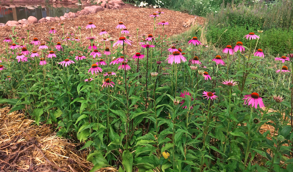
It seems that a big part of Echinacea’s immune stimulation is directly attributable to endophytic (and surface) bacteria. More specifically, bacterial lipopolysaccharide (LPS) and lipoproteins are the culprits. These are actually not the secondary metabolites I’ve been blabbing about but are structural components of bacterial membranes. Anyway, my first thought was “WTH????” after reading that bacterial stuff is largely responsible for Echinacea’s immune activation. And, Echinacea is loaded with endophytes. One study alone isolated over 150 endophytic bacterial species (1), and this isn’t even considering how many are there but not able to be grown in culture or how many fungal species there are.
How did folks decide that bacteria are important for Echinacea’s immune stimulation? One way was to grow Echinacea plants from seeds that had been sterilized of any endophytes. Ethanolic extracts from the resultant endophyte-free Echinacea plants were not active in stimulating macrophage activity, while the control plants containing bacterial endophytes were active (2). Macrophages are important cells in our innate immune response, and the ability to activate macrophages in culture is a common way to assess immune stimulation by plants, drugs, etc. When I first read this, I figured it didn’t eliminate Echinacea polysaccharides as important contributors to the plant’s immune stimulating activity, since the extracts studied were made with ethanol and largely lacked plant polysaccharides. And, I stuck my tongue out and blew a raspberry in the general direction of the researchers (North Carolina).
But, research with polysaccharides extracted from Echinacea also found that inactivating bacterial LPS and lipoproteins in the extracts destroyed most, though not all, of the immune stimulating activity (3). This was true not just in the standard macrophage assay in a dish, but in vivo as well (3). Even in column purified polysaccharide preparations, bacterial lipoproteins are present (3), and it really doesn’t take much of either LPS or lipoprotein to trigger an immune response. Endophytes were sources of LPS and lipoproteins, though some may come from surface-contaminating bacteria as well (3). And these observations weren’t limited to Echinacea…similar results were seen with Black Walnut, American Ginseng, Alfalfa and others (3). Gah!!!
More along these lines… When Echinacea preparations from different growers were assessed, they varied in levels of immune stimulation by about 100-fold, and this variation in activity was closely tied to the level of bacterial LPS and lipoproteins in the preparations rather than, for instance, the method of plant processing (4). And it’s not just any old bacterium that is responsible. Even though overall numbers of endophytic bacteria in Echinacea preparations correlated with how robust the immune stimulation was, particular bugs, such as Proteobacteria, were linked with the extent of immune stimulation (1, 2).
Alas, all is not lost for the polysaccharides. There is still residual immune stimulation when the LPS/lipoproteins are inactivated that could be due to the polysaccharides. Also, components of the immune system not evaluated in these various studies may be triggered by the polysaccharides. In any event, the story is maybe a bit more complicated than what we see in the standard herb book.
Aside from immune stimulating activity, Echinacea is also used for immune modulating effects, reducing inflammation for example. Immune modulation by Echinacea is due partly to plant alkylamides, whose effects are mediated in part via cannabinoid receptors on immune cells (5). Alkylamides, for example, suppress the production of pro-inflammatory molecules such as Tumor Necrosis Factor ? (TNF-?) by macrophages. As mentioned, it’s possible to grow plants lacking endophytes. Endophyte-free Echinacea plants had lower levels of alkylamides compared to plants innoculated with bacterial (6) or fungal (7) endophytes isolated from other Echinacea plants. In other words, the presence of certain endophytes increases alkylamide levels in Echinacea; yet another way endophytes significantly influence this commonly used and much valued medicine.
Ashwagandha
OK, so Echinacea is more that what it seems at first glance. How about another extensively used medicinal plant (and one of my favorites), Ashwagandha?
As you can probably guess, given the topic of this class and article, endophytes are at it again…

Withanolides are steroidal lactones and are the best studied constituents of Ashwagandha. Withanolides have a broad range of activities: Anti-inflammatory, anti-oxidant, anti-tumor, radioprotective, anti-bacterial, immunomodulating, liver protective, anti-rheumatic, anti-anxiety, etc etc etc….(8). Both bacterial and fungal endophytes significantly increase withanolide production in Ashwagandha root and other parts of the plant (9, 10) by activating plant genes involved in withanolide synthesis (9). For those who care, withaferin A, withanolide A, withanolide B and 12-deoxy-withstramonolide all went up. Withafarin A was not detectable in Ashwagandha roots lacking endophytes, but was found in the roots in significant amounts in plants innoculated with certain endophytes (9). For other microbiology geeks out there, some of the responsible bugs found in roots, leaf and stem included the bacteria Bacillus, Staphylococcus and Pseudomonas and the fungi Aspergillus and Penicillum (9).
What is really cool is that some endophytes, like the fungus Taleromyces pinophilus, can make withanolides on their own, and production by fungal isolates was more robust than by the plant itself (11)! How is it that a fungus can make the same “plant medicine” as the plant itself? Not clear. Possibly, it’s an example of “horizontal” gene transfer between species. (As opposed to “vertical” gene transfer, which is inheritance from parent(s) to offspring.) Somehow the fungus seems to have picked up Ashwagandha genes.
Ginkgo
I’ve been on Ginkgo since getting kicked in the head by a spazz of a training partner. Between that and dementia on both sides of the family, taking Ginkgo seems like a good idea. Ginkgo leaf extracts improve cognitive function via better cerebral blood flow as well as by acting as a brain anti-oxidant: Rust-o-leum for the noodle, as it were. Ginkgo also improves circulation to other parts of the body, including the reproductive system, and has protective effects on the cardiovascular system. Ginkgo may even have some cancer preventative effects, showing activity in cell culture and animal models (12). Some well known Ginkgo metabolites contributing to its medicinal effects include terpenoids such as the creatively named bilobalide and ginkgolides, along with multiple flavonoids.

Ginkgo’s resident critters represent the various ways that endophytes influence the secondary metabolites found in plants. For example, the fungal entophyte Pestalotiopsis uvicola can make bilobalide independent of the plant (13). Another fungal endophyte, Fusarium oxysporum, can synthesize ginkgolide B (14), while Aspergillus species synthesize Ginkgo flavonoids (15). Other fungal endophytes can increase Ginkgo’s own production of flavonoids (16). And then there’s a fungal endophyte, Chaetomium globosum, that produces anti-tumor metabolites called chaetoglobosins that the Ginkgo itself doesn’t make (17).
I think this is interesting in and of itself simply from a nerd’s point of view. But, just think…if fungi can produce these things in culture, maybe nutraceutical companies will start leaving the plants alone and work with microbial cultures to produce medicinal constituents. Less pressure on the plant. I know, you’re grumbling right now that plant medicine is more than just a few isolated chemicals. I agree fully. But, hey, if a company is obsessed with the “active principles”, let ‘em produce them in a dish instead of razing the countryside.
Tulsi
On to Tulsi (aka Holy Basil), the “Queen of Herbs” in Ayurveda. Among other uses, Tulsi has traditionally been turned to for diabetes. Tulsi leaf extracts reduce blood sugar in part by inhibiting alpha – amylase, a key enzyme in breaking down starch and glycogen into simple sugars. I wonder how much of this activity is due to the plant itself versus resident endophytes, given that metabolites produced by the fungal endophytes Alternaria, Colletotrichum, Diaporthe and Trichoderma isolated from Tulsi leaf and twig strongly inhibited not only alpha – amylase but also alpha – glucosidase (18), which breaks starch and disaccharides down into glucose. In addition, the endophyte metabolites inhibited aldose reductase (18), an enzyme in glucose metabolism that contributes to diabetic complications in the eyes, kidneys and elsewhere.
Multiple studies show the potential for Tulsi in cancer prevention (19), and the fungal Alternaria, Phoma and Exserohilum may contribute by synthesizing metabolites able to kill cancer cells (20). At least, cells in a dish, keeping in mind that this doesn’t always translate to efficacy in an actual person.
Tulsi is useful for dealing with infections of various ilk, being broadly active against pathogenic microbes. It’s possible that its endophytic hitchhikers play some role in this. For example, the fungal endophytes mentioned above also have activity against several pathogenic bacteria (20), and other endophytes from Tulsi can stimulate the activity of neutrophils (21), which fight infection as part of the innate immune response.
Tulsi has been used also for its organ-protective effects: Liver, kidneys, brain and such. Crude fractions of the fungus Paecilomyces isolated from Tulsi protected the liver from oxidative damage in experimental models (22), so maybe endophytes contribute to Tulsi’s protective effects as well. To be clear, how involved endophytes are in these various activities of Tulsi — enzyme-inhibition, cancer prevention, resolving infection, organ health — presumably depends on the significance of the impact on the secondary metabolites present within the plant. So, it’s simply not yet clear.
One more note on Tulsi. It has a very characteristic scent. Whether it’s the Krishna, Rana or Vana variety, it’s still identifiable as Tulsi even though the essential oil chemistry differs somewhat among these varieties. Tulsi’s endophytes may influence the characteristics and strength of the scent. This goes beyond a pleasant scent, as plant volatile oils are often a significant part of the medicine. The fungal endophyte Macrophomina phaseolina is abundant in Tulsi leaf and produces the volatile oil 2H- pyran-2-one, 5,6-dihydro-6-pentyl (24). (Say that 5 times fast.) That mouthful is otherwise known as 10C-Massia lactone, which has a creamy, sweet, fruity scent (25). Bacterial endophytes, including Bacillus subtilis, can enhance both plant volatile oil production as well as plant size (23). Perhaps something to be taken advantage of in large scale essential oil production…fewer plants for the same amount of oil.
Red Sage
Red Sage root (Danshen) is valued in modern Chinese Medicine for cardiovascular benefits. Red Sage is immune modulating, anti-oxidant (really, is there a medicinal plant that isn’t?), anti-tumor, anti-inflammatory, anti-microbial…the usual collection of “antis”. Metabolites characterized so far include flavonoids, terpenoids, salvianolic acids and other phenolics. The most well characterized are diterpenoids known as tanshinones, which contribute to all of the activities noted above (26).
Chaetomium globosum, a fungal isolate from Red Sage root (and also of Ginkgo fame!), can significantly increase tanshinone production in the root (27). In the absence of this endophyte, tanshinone levels were quite low (27). Levels of salvainolics acid were also bumped up and root and overall plant size were increased as well (27). Salvainolic acids have multiple ways of protecting the heart and vasculature from damage.
Other endophytes from Red Sage synthesize flavonoids, saponins, steroids, tannins, alkaloids, terpenoids and phenolics, including salvionolic acid C, that the plant also makes (28, 30); another case of the bugs making some of the same medicines the plant makes. Again, an increase in plant size was seen in response to certain endophytes (29), and some endophytes could even synthesize metabolites — cardiac glycosides and anthroquinones — not otherwise found in the plant (28). Cardiac glycosides have obvious relevance to the cardiovascular system, and some anthroquinones may impact blood vessel contractility. Interesting, given the use of Red Sage for the cardiovascular system.
Guduchi (Tinospora cordifolia)
Based on the most of the plants in this article, you may be guessing that a lot of the research on endophytes in medicinal plants is being carried out in Asia, and you’d be correct. Along those lines, there is a growing pile of info on how endophytes impact Guduchi, another valued medicine from the Ayurvedic tradition. Yet another multifaceted herbal medicine, Guduchi is commonly used, among other things, for urinary issues ranging from gravel and stones to bladder weakness. TMI warning. When on Guduchi, even for less than a week, I only had to get up to pee once at night instead of the usual 2-3 times. (Why am I not still on it ????)
Guduchi is also used for gout, and it’s been a component in my formulas the few times I’ve worked with this. Gout is the, at times excruciating, accumulation of uric acid in joints, particularly in but not limited to the big toe. Gout sucks. Xanathine oxidase is an enzyme involved in uric acid production. Several endophytes isolated from Guduchi make inhibitors of xanathine oxidase, perhaps contributing to Guduchi’s benefits in gout (31). So, again, a case where the endophytes may be the source of some of the activity of our herbal medicines.
Guduchi extracts have shown various anti-tumor activities in preclinical trials (ie. cell culture and animal models). A fungal endophyte isolated from Guduchi, Fusarium culmorum, produces Taxol (32), one of the most widely used chemotherapy drugs. Another endophyte, Cladosporium velox, produces a bunch of polyphenols — gallic acid, catechin, chlorogenic acid, epicatechin, caffiec acid, coumaric acid, ellagic acid and others — with with genoprotective and anti-oxidant effects (33). Genoprotective agents reduce DNA mutations and that’s relevant to cancer because, with only a handful of exceptions, cancer has gene mutations as it’s root.
Others…

Not to be left out, St. John’s Wort has a fungal endophyte (Thielavia subthermophila) that makes hypericin (34), while root-associated mycorrhial fungi can stimulate increased production of both hypericin and pseudohypericin by St. John’s Wort itself (35).
Those of you who use Milk Thistle may be familiar with silymarin, a seed extract containing over a half dozen flavinoligan compounds and used for hepatoprotective effects. A few of these flavinoligans — Silybin A, silybin B, and isosilybin A — can be made by a fungal endophyte, Aspergillus iizukae, that lives in the leaves of the plant (36).
Ginsenosides are steroidal saponins in Panax Ginseng that contribute significantly to the many uses of this vastly popular plant medicine. So far we know that two of these, ginsenoside Rb2 and ginsenoside Rc, can be made by fungal endophytes from the root (37).
And there are many, many more examples of how our favorite medicines may be slightly more than just “herbal” medicines. To summarize, the various outcomes of endophyte-plant interactions on secondary metabolite production include: (i) endophytes making something that the plant doesn’t make, (ii) endophyte stimulating the plant to make something (or to make something at higher levels) and (iii) both endophyte and plant making the same metabolites. This last one seems the most weird. What’s the benefit to endophyte or plant when both are making the same metabolites? Who knows….
Anyway, what’s the point of all of this? A big part of it is me geeking out on some cool microbiology. Research into endophytes and medicinal plants has grown quite a bit over the past 10 years. More practically, there’s the thought that adding certain endophytes to farmed medicinal plants may improve plant size and yields, cutting down on environmental impact and, hopefully, helping smaller growers make a better living. That said, there are potential issues with sprinkling endophytes willy nilly on plants. For instance, what’s friendly to one plant species may not be to another.
As mentioned earlier, crude extracts are where it’s at for many herbalists, including myself. Most standardized extracts seem to be bullshit, as additional “active principles” keep being found for various plants that are marketed as standardized extracts (eg. Turmeric, St. John’s Wort, others). You may disagree. You may not. And, isolated components, such as curcumins, lack the full effect of the crude extract and have more side effects. But, I’ll mention again that I do like the idea of having alternate sources of secondary metabolites, cultured endophytes, so that nutraceutical companies are perhaps less likely to wipe out plants x, y or z to obtain such metabolites for their products.
~~~
Content © Dr. Anna Marija Helt, Osadha Natural Health, LLC. Permission to republish any of the articles or videos in full or in part online or in print must be granted by the author in writing.
The articles and videos on this website for educational purposes only & have not been evaluated by the Food and Drug Administration. This information is not intended to diagnose, treat, cure, or prevent any disease or to substitute for advice from a licensed healthcare provider.
References
1. Haron, MH, et al (2014) Immune enhancing Echinacea bacterial endophytes. Planta Med. 80 – PP15. https://www.thieme-connect.com/products/ejournals/abstract/10.1055/s-0034-1382710
2. Todd, DA, et al (2015) Ethanolic Echinacea purpurea extracts contain a mixture of cytokine-suppressive and cytokine-inducing compounds, including some that originate from endophytic bacteria. PLOS One. 10(5): e0124276. https://journals.plos.org/plosone/article?id=10.1371/journal.pone.0124276
3. Pugh, ND, et al (2008) The majority of in vitro macrophage activation exhibited by extracts of some immune enhancing botanicals is due to bacterial lipoproteins and lipopolysaccharides. Int Immunopharmacol. 8(7):1023-32. https://www.ncbi.nlm.nih.gov/pmc/articles/PMC2467439/
4. Tamta, H, et al (2008) Varability in in vitro macrophage activation by commercially diverse bulk Echinacea plant material is due predominantly to bacterial lipoproteins and lipopolysaccharides. J Agric Food Chem. 56(22):10552-10556. https://www.ncbi.nlm.nih.gov/pmc/articles/PMC2670557/pdf/nihms91114.pdf
5. Raduner, S, et al (2006) Aklylamides from Echinacea are a new class of cannabinomimetics: CAnnabinoid type 2 receptor-dependent and -independent immunomodulatory effects. J Biol Chem. 281:14192-06. http://www.jbc.org/content/281/20/14192.full.html
6. Maggini, V, et al (2017) Plant-endophytes interaction influences the secondary metabolism in Echinacea purpurea (L.) Moench: an in vitro model. Scientific Rep. 7:16924. https://www.nature.com/articles/s41598-017-17110-w
7. Gualandi, RJ Jr. (2010) Fungal endophytes enhance growth and production of natural products in Echinacea purpurea (Moench.). ” Master’s Thesis, University of Tennessee. https://trace.tennessee.edu/utk_gradthes/713/
8. Budhiraja, RD, et al (2000) Biological activity of withanolides. J Sci Indust Res. 59:904-11. buhttp://nopr.niscair.res.in/bitstream/123456789/26628/1/JSIR%2059(11)%20904-911.pdf
9. Pandey, SS, et al (2018) Endophytes of Withania somnifera modulate in planta content and the site of withanolide biosynthesis. Scientific Reports. 8:5450. https://www.researchgate.net/publication/324173010_Endophytes_of_Withania_somnifera_modulate_in_planta_content_and_the_site_of_withanolide_biosynthesis
10. Mishra, A et al (2018) Bacterial endophytes modulates the withanolide biosynthetic pathway and physiological performance in Withania somnifera under biotic stress. Microbiol Res. Jul – 212-213:17-28. https://www.ncbi.nlm.nih.gov/pubmed/29853165
11. Sathiyabama, M & R Parthasarathy (2018) Withanolide production by fungal endophyte isolated from Withania somnifera. Nat Prod Res. 32(13):1573-77. https://www.ncbi.nlm.nih.gov/pubmed/29034745
12. DeFeudis, FV, et al (2003) Ginkgo biloba extracts and cancer: A research area in it’s infancy. Fundam Clin Pharmacol. 17(4):45-17. https://www.ncbi.nlm.nih.gov/pubmed/12914542
13. Qian, Y-X, et al (2016) A bilobalide-producing endophytic fungus, Pestalotiopsis uvicola from medicinal plant Ginkgo biloba. Curr Microbiol. 73(2):280-6. https://link.springer.com/article/10.1007/s00284-016-1060-6
14. Cui, Y, et al (2012) Ginkgolide B produced endophytic fungus (Fusarium oxysporum) isolated from Ginkgo biloba. Fitoterapia 83(5):913-20. https://www.sciencedirect.com/science/article/pii/S0367326X12001165
15. Qiu, M, et al (2010) Isolation and identification of two flavonoid-producing endophytic fungi from Ginkgo biloba L. Annal Microbiol. 60(1):143-150. https://www.researchgate.net/publication/225651927_Isolation_and_identification_of_two_flavonoid-producing_endophytic_fungi_from_Ginkgo_biloba_L
16. Hao, G, et al (2010) Fungal endophytes-induced abscisic acid is required for flavonoid accumulation in suspension cells of Ginkgo biloba. Biotechnol Let. 32(2): 305–314. https://link.springer.com/article/10.1007/s10529-009-0139-6
17. Li, H, et al (2014) Chaetoglobosins from Chaetomium globosum, an endophytic fungus in Ginkgo biloba, and their phytotoxic and cytotoxic activities. J Agric Food Chem. 62(17):3734-41. https://www.researchgate.net/publication/261441769_Chaetoglobosins_from_Chaetomium_globosum_an_Endophytic_Fungus_in_Ginkgo_biloba_and_Their_Phytotoxic_and_Cytotoxic_Activities
18. Pavritha, N, et al (2014) In-vitro Studies on ?-Amylase, ?-Glucosidase and Aldose Reductase Inhibitors found in Endophytic Fungi Isolated from Ocimum sanctum. Curr Enz Inhibition. 10:129-36. https://www.researchgate.net/publication/269694533_In-vitro_Studies_on_a-Amylase_a-Glucosidase_and_Aldose_Reductase_Inhibi-_tors_found_in_Endophytic_Fungi_Isolated_from_Ocimum_sanctum
19. Baliga, MS (2013) Ocimum sanctum L (Holy Basil or Tulsi) and its phytochemicals in the prevention and treatment of cancer. Nutr Cancer. 65 Suppl 1:26-35. https://www.ncbi.nlm.nih.gov/pubmed/23682780
20. Bhagat, J, et al (2010) Molecular and functional characterization of endophytic fungi from traditional medicinal plants. World J Microbiol Biotechnol. 28(3):963-71.https://www.ncbi.nlm.nih.gov/pubmed/22805817
21. Madagundi, S, et al (2013) Free radical scavenging and in vitro immunomodulatory activites of endophytic fungi of Ocimum sanctum Linn. Farmacia. 61(2):330. https://www.researchgate.net/publication/261418046_FREE_RADICAL_SCAVENGING_AND_IN_VITRO_IMMUNOMODULATORY_ACTIVITIES_OF_ENDOPHYTIC_FUNGI_OF_OCIMUM_SANCTUM_LINN
22. Shukla, ST, et al (2012) Hepatoprotective and antioxidant activities of crude fractions of endophytic fungi of Ocimum sanctum Linn. Oriental Pharm Exp Med. 12(2):81-91. https://www.researchgate.net/publication/257805712_Hepatoprotective_and_antioxidant_activities_of_crude_fractions_of_endophytic_fungi_of_Ocimum_sanctum_Linn_in_rats
23. Tiwari, R (2010) Endophytic Bacteria from Ocimum sanctum and Their Yield Enhancing Capabilities. Curr Microbiol. 60(3):167-71. https://www.researchgate.net/publication/26892447_Endophytic_Bacteria_from_Ocimum_sanctum_and_Their_Yield_Enhancing_Capabilities
24. Chowdhary K, Kaushik N (2015) Fungal Endophyte Diversity and Bioactivity in the Indian Medicinal Plant Ocimum sanctum Linn. PLoS ONE 10(11): e0141444. 90 https://journals.plos.org/plosone/article?id=10.1371/journal.pone.0141444
25. http://www.thegoodscentscompany.com
26. Wang, B-Q (2010) Salvia miltiorrhiza: Chemical and pharmacological review of a medicinal plant. J Med Plant Res. 4(25):2813-20.http://www.academicjournals.org/app/webroot/article/article1380714402_Wang.pdf
27. Zhai, X, et al (2018) Endophyte Chaetomium globosumD38 Promotes Bioactive Constituents Accumulation and Root Production in Salvia miltiorrhiza. Front Microbiol.
8:2694. https://www.frontiersin.org/articles/10.3389/fmicb.2017.02694/full
28. Li, Y, et al (2015) The endophytic fungi of Salvia miltiorrhiza Bge.f. alba are a potential source of natural antioxidants. Bot Stud. 56: 5 https://www.ncbi.nlm.nih.gov/pmc/articles/PMC5430307/
29. Zhou, LS, et al (2018) The Plant Growth-Promoting Fungus (PGPF) Alternaria sp. A13 Markedly Enhances Salvia miltiorrhiza Root Growth and Active Ingredient Accumulation under Greenhouse and Field Conditions. Int J Mol Sci. 19(1):270. https://www.ncbi.nlm.nih.gov/pmc/articles/PMC5796216/
30. Li, X, et al (2016) Phoma glomerata D14: An Endophytic Fungus from Salvia miltiorrhiza That Produces Salvianolic Acid C. Curr Microbiol. 73(1):31-7. https://www.researchgate.net/publication/298740061_Phoma_glomerata_D14_An_Endophytic_Fungus_from_Salvia_miltiorrhiza_That_Produces_Salvianolic_Acid_C
31. Kapoor, N & S Saxena. (2018) Endophytic fungi of Tinospora cordifolia with anti-gout properties. 3 Biotech. 8(6):264. https://www.researchgate.net/publication/325254828_Endophytic_fungi_of_Tinospora_cordifolia_with_anti-gout_properties
32. Visalakchi, S, et al (2010) Taxol producing endophytic fungus Fusarium culmorum SVJM072 from medicinal plant of Tinospora cordifolia – a first report. J Biotechnol. 150:425-425. https://www.researchgate.net/publication/241082625_Taxol_producing_endophytic_fungus_Fusarium_culmorum_SVJM072_from_medicinal_plant_of_Tinospora_cordifolia_-_a_first_report
33. Singh, B, et al (2016) Antioxidant and in vivo genoprotective effects of phenolic compounds identified from an endophytic Cladosporium velox and their relationship with its host plant Tinospora cordifolia. J Ethnopharmacol. 194:450-6. https://www.researchgate.net/publication/308993108_Antioxidant_and_in_vivo_genoprotective_effects_of_phenolic_compounds_identified_from_an_endophytic_Cladosporium_velox_and_their_relationship_with_its_host_plant_Tinospora_cordifolia
34. Kusari, S, et al (2008) J Nat Prod. 71(2):159-162. An endophytic fungus from Hypericum perforatum that produces hypericin. https://www.ncbi.nlm.nih.gov/pubmed/18220354
35. Zubek, S, et al (2012) Hypericin and pseudohypericin concentrations of a valuable medicinal plant Hypericum perforatum L. are enhanced by arbuscular mycorrhizal fungi. Mycorrhiza. 22(2):149–156. https://www.researchgate.net/publication/51178213_Hypericin_and_pseudohypericin_concentrations_of_a_valuable_medicinal_plant_Hypericum_perforatum_L_are_enhanced_by_arbuscular_mycorrhizal_fungi
36.El-Elimat, T, et al (2014) Flavonolignans from Aspergillus iizukae, a fungal endophyte of milk thistle (Silybum marianum). J Nat Prod 77(2):193-9. https://libres.uncg.edu/ir/uncg/f/H_Raja_Flavonolignans_2014.pdf
37. Wu, H, et al (2013) Diversity of endophytic fungi from roots of Panax ginseng and their saponin yield capacities
SpringerPlus. 2:107. https://www.researchgate.net/publication/236095071_Diversity_of_endophytic_fungi_from_roots_of_Panax_ginseng_and_their_saponin_yield_capacities
Defeudis, FV (2002) Bilobalide and neuroprotection. Pharmacol. Res. 46(6):565-8. https://www.sciencedirect.com/science/article/abs/pii/S1043661802002335.